|
Tel: 888 374 9411 Fax: 888 374 9412 Email: dentistry.specialists@dsilc.com |
|
|
Tel: 888 374 9411 Fax: 888 374 9412 Email: dentistry.specialists@dsilc.com |
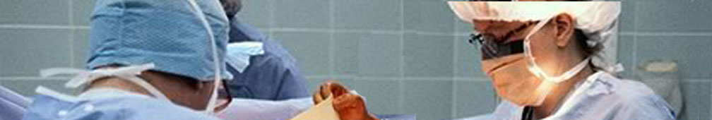
DSI uses digital X-Rays to provide even better dental care and more accurate diagnoses. The process of digitally capturing an image is much faster, safer and more comfortable for the patient.
X-ray images, also called dental radiographs, are among the most valuable tools a dentist has for keeping your mouth and teeth healthy. By understanding what the structures of the mouth look like normally on an X-ray film, dentists can diagnose problems in the teeth and jaws. For adults, radiographs can:
We place a small sensor in your mouth. The sensor is connected to a computer by a thin wire. Next, an X-ray beam is sent through your teeth and into the sensor, which records the image of your teeth and sends it to the computer. The sensor can then be repositioned to photograph other sections of your teeth.
The digital dental X-ray system is more sensitive than dental X-ray film systems, so your exposure to X-rays is cut by as much as 90 percent. The large, color-enhanced images let you see what your dentist sees, so it's easier for you to understand how your dentist will treat your teeth. Your fees don't include payment for photo chemicals, film, processing or film storage. Used photo chemicals and film are not polluting the environment. Your dental checkups take less time, and it's fun to watch this system work! Most patients are amazed.
Perhaps the future of dental X-Ray diagnostics lies within the technology known as 3D Conebeam CT (Computed Tomography).
Computed tomography (CT) is currently most often made with an array of (e.g. 64) linear detectors, employing reconstruction algorithms that are a compromise between 2D and 3D geometry. Among the advantages of this technique are superior resolution and lower radiation dose compared with prior technologies. The images produced by these technologies also reveal information that traditional technologies could not—such as the ability to rotate images, see the relationship of nerves and tissue to teeth, and the ability to significantly magnify images for greater detail.
SI is investing in this technology to provide the most accurate and advanced diagnostic tools for patient care. If appropriate, the patient will be sent to our central facility or to a partner facility for diagnostic procedures.